


Translational Oncology
Research group Translational Oncology
Magnetic Resonance Spectroscopy (MRS) is an MR-based technology that allows non-invasive examination of biochemical changes in tumors. It provides in vivo metabolic information that is not accessible by any other means using currently available experimental methods. The physical instrumentation, pulse sequences, and postprocessing methods that involve complex data analysis algorithms are our area of expertise in brain tumors. X-nuclear MRS has been used at our institution to measure various parameters including grading and staging of brain tumors with the identification of tumor specific mutations, pharmacodynamics (when a drug contains MRS-detectable nuclei such as 19F) in trials of new oncology drugs and early response of brain tumors to treatment and dietary interventions. In new projects we apply our broad knowledge to lymphoma patients with superficial lymph node manifestations and explore glutamatergic mechanisms in glioma patients.
Multi-parametric qMRI refers to the acquisition of different quantitative parametric MR maps of the brain in clinical acquisition times. The obtained parameters provide information on the underlying micro-structural organization of the examined tissue. As opposed to weighted images, they constitute measures in physical units which give them the potential to obtain measures with a high degree of reproducibility across time points, scanner manufacturers, scanner models, and sites. The obtained imaging biomarkers can serve as ground truth for AI image postprocessing solutions and multiply their potential in brain tumor research.
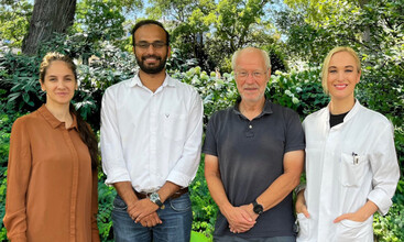
The group leader Katharina Wenger-Alakmeh is a board-certified radiologist and assistant professor (Habilitation) at the Institute of Neuroradiology with prior clinical experience in Neurology/Neurooncology. Following her time as a Junior Clinician Scientist in 2016 (FFF), she became a fellow of the Mildred Scheel-Clinician Scientist program (German Cancer Aid) in October 2021. In addition, she has received funding from the Else Kröner-Fresenius-Stiftung. Throughout past projects she has worked with multidisciplinary groups of fellow researchers with the aim of translating innovative technologies into the clinical setting. She has long-standing experience in MR-based metabolic tumor imaging working with patients and small animals.
Seyma Alcicek is an MD-PhD with prior experience in nuclear medicine which allowed her to gain insights into several oncological imaging techniques. During her PhD studies as a Marie Slodowska-Curie early-stage researcher at Jagiellonian University, she focused on practical applications of novel spectroscopic methods for the realization of their use in clinical studies. She became a fellow of the Mildred Scheel-Medical Scientist program in April 2022. Her research focuses on understanding the metabolic features of various tumor types in a non-invasive manner using in vivo magnetic resonance spectroscopy and improving the feasibility of this technique for clinical application.
Dennis C. Thomas is an MR Imaging scientist with expertise in the field of quantitative imaging. He pursued his PhD studies as a Marie Slodowska-Curie early-stage researcher at Forschungszentrum Jülich and focused on novel quantitative MRI techniques to develop new biomarkers of the diseased brain. His doctoral work was part of the EU-funded “project B-Q Minded” which focused on the advancement of Q-MRI techniques, especially in its application in the clinic. Dennis’ current research interests include applications of robust qMRI techniques in the clinic and the development of reliable and reproducible MRI biomarkers of brain tumors.
The group is advised by Dr. rer. nat. Ulrich Pilatus, an MR Physicist focused on metabolic imaging of oncological and neurological diseases with prior international positions (University of Maryland, Baltimore County (USA); Weizmann Institute of Science (Israel)) and over 20 years of experience in the field.
Methods
X‑nuclei (1H, 31P, 19F) magnetic resonance imaging (MRI) on 3T clinical and 7T preclinical scanners using volume and surface coils
Quantitative MRI: T1 and T2* relaxometry (qT1 & qT2*) and Water content mapping (PD)