Research

Cognitiv


Cognitive Neuroscience

The Cognitive Neuroscience group has several research foci. Our main aim is discovering the temporal characteristics of interareal interactions in brain networks that may offer additional insight into brain function compared to the study of spatial characteristics of brain activity alone. Particularly higher cognitive functions like speech, language, and executive control require the interaction between multiple brain regions. We study how complex behavior is related to the spatio-temporal organization of brain networks with a special emphasis on hemispheric lateralization, sensorimotor integration, and circadian rhythms. The projects use data from functional and structural magnetic resonance imaging, magnetoencephalography, electrocorticography, intracranial electroencephalography, and interventions like direct cortical or subcortical electrical stimulation. Studies on people who stutter and patients with Parkinson’s disease, brain tumor, or epilepsy complement investigations in healthy participants. If you would like to learn more about our work and team, please click here.


Members

Publications

Movement2


Movement Disorders

The main research focus of our group is the investigation of brain microstructure (and function) in various movement disorders, such as Parkinson’s disease, atypical Parkinsonism and dystonia. To this end, we use multimodal quantitative imaging techniques like T1-, T2-, T2*- and proton density mapping which allow the assessment of important tissue properties as, for example, the iron and water content and the degree of myelination.  With these methods we seek to define disease specific patterns of tissue change and to link them with the patients’ clinical phenotype. One goal is to improve our general pathomechanistic understanding of the disease and to entangle the structural correlates of specific motor or non-motor symptoms frequently observed in movement disorders (e.g. akinesia or cognitive decline). A second goal is the development of image based biomarkers which may be used for clinical differential diagnosis of movement disorders and, also, for the monitoring of disease progression in longitudinal clinical studies. Another important thematic focus is the development and exploration of novel imaging techniques, e.g. methods that facilitate the acquisition of quantitative MRI parameters in the clinical setting. This takes place in close cooperation with the core structure of the BIC. 


Members

Publications


Structural Imaging Group

Quantitative MRI (qMRI) techniques measure actual tissue parameters and allow for the assessment of the microstructural composition even in tissue which appears normal on conventional MRI (Deichmann and Gracien (2018), in: Quantitative MRI of the brain: Principles of physical measurement).

A main research focus of our group (Principal investigators PD Dr. Alexander Seiler and Prof. Dr. René-Maxime Gracien) is the investigation of diffuse changes in tissue composition in patients with neuroinflammatory diseases with quantitative MRI (qMRI) and diffusion tensor imaging techniques and of the relationship of these abnormalities with the clinical and cognitive status. Additionally, we evaluate the interaction between functional and structural changes in Multiple Sclerosis (MS) with a combined fMRI (functional MRI) and qMRI approach. We are furthermore interested in the development and validation of qMRI techniques (Gracien et al., 2019, Neuroimage) and synthetic contrasts (Gracien et al., 2019, Magn Reson Imaging) in cooperation with the physicists of our Brain Imaging Center (BIC, especially Prof. Ralf Deichmann).
As an example, we were able to demonstrate that cortical T1 and proton density (PD) increases are already present in early MS stages, reflecting changes in cortical architecture (Gracien et al., 2016, Eur Radiol). Another study characterized cortical pathology by applying a multi-parametric qMRI protocol including T1, PD and magnetization transfer ratio (MTR) mapping in a relapsing-remitting MS (RRMS) patient group, which covered a broad range of disability severity and disease duration, and revealed a relationship between cortical T1 relaxation time and the clinical status (Gracien et al., 2016, J Magn Reson Imaging). In secondary progressive MS (SPMS) global neurodegeneration gains significance which cannot be directly quantified with conventional MRI. A previous study indicated that qMRI methods might be promising candidates for the assessment of neurodegeneration in relationship to the degree of disability in SPMS (Gracien et al., 2016, PloS One). Furthermore, we demonstrated spatially inhomogeneous cortical changes of qMRI parameters in RRMS with parameter-specific patterns (van Wijnen, ..., Gracien, 2019, Eur Radiol). In this study cortical T2 and T2* values were correlated with the cognitive status. Our results indicated that the patient’s pattern of cognitive changes might depend on the distribution of cortical damage. Furthermore, we investigated tissue remodeling caused by natural ageing (Gracien, Nürnberger et al., 2017, Eur Radiol) and neurodegeneration in Parkinson’s disease with T1 relaxometry (Nürnberger, Gracien et al., 2017, Neuroimage: Clin).
In addition, we develop methods for the improved visualization of epileptogenic lesions in close cooperation with the BIC core structure (Prof. R. Deichmann, Dr. U. Nöth), Prof. M. Wagner (neuroradiologist at Betanien hospital) and the Institute of Neuroradiology (Prof. E. Hattingen) at Goethe University and assess tissue properties outside of these lesions in patients with epilepsy with qMRI techniques.
Figure website a seiler

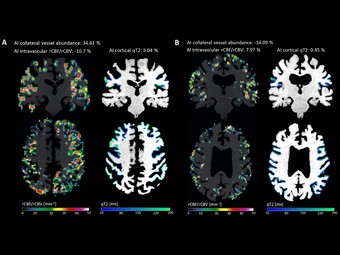
Regarding cerebrovascular diseases, one of our main research interests is investigating pathological changes of brain energy metabolism and cerebral microstructure in patients with chronic cerebral hypoperfusion. Due to the prognostic significance of altered brain oxygen metabolism concerning the occurrence of permanent ischemic tissue damage and the risk of (recurrent) ischemic stroke, the MRI-based characterization of tissue oxygen metabolism in patients with cerebral large-artery steno-occlusive disease has been one of our major research activities in recent years (Seiler et al., 2012, Stroke, Seiler et al., 2016, PLOS ONE, Seiler et al., 2019, Stroke), which we are further pursuing for continuous improvement and validation of oxygenation-sensitive MR imaging techniques. Furthermore, were are interested in hypoperfusion-associated tissue damage on the microstructural level and its potential link to cognitive impairment (Seiler et al., 2018, Am J Neuroradiol, Seiler et al., 2020, J Cereb Blood Flow Metab). Apart from the investigation of chronic hemodynamic impairment, part of our research focuses on methodological approaches for the assessment of collateral supply (Seiler et al., 2019, J Cereb Blood Flow Metab) and the metabolic description of tissue-at-risk in acute ischemic stroke (Seiler et al., 2017, Stroke, Seiler et al., 2019, Clin Neuroradiol, Seiler et al., 2019, J Cereb Blood Flow Metab). Further projects involve the evaluation of potential imaging biomarkers of cognitive decline in patients with small vessel disease as well as lesional and perilesional tissue characterization in patients with intracerebral hemorrhage.

Cooperations:
-Prof. R. Deichmann, Head of the core structure of the BIC
-PD S. Baudrexel, Movement Disorders Group
-PD J. Klein, University of Oxford
-Prof. E. Hattingen, Institute of Neuroradiology
-Prof. M. Wagner, Betanien hospital
-Prof. C. Förch, Multiple Sclerosis Group
-Prof. F. Rosenow, Epilepsy Center Frankfurt
-Prof. S. Knake, Epilepsy Center Marburg

Members

Publications

Epilepsy

The Epilepsy Imaging Group is a new research group associated with the Epilepsy Center Rhein-Main.
Our target is the multimodal assessment of neural activity in epilepsy and health. We combine structural MR imaging with functional methods, especially simultaneous EEG-fMRI and MEG. Combining these methods with intracranial EEG data, we aim to identify epileptogenic networks in focal and generalized epilepsies. Analysis tools comprise traditional generalized linear models and connectivity analyses as well as time-frequency and information-theoretic techniques.
In many epilepsy patients, conventional MRI is not able to identify structural anomalies, even though surface EEG may clearly show a focus of increased electrical excitability. We therefore apply state-of-the-art quantitative MRI techniques to improve the detection of presumed structural lesions.
Projects are carried out in collaboration with the Departments of Neuroradiology and Neurosurgery and the MEG laboratory of the Goethe University.

Members

Publications